

Traditionally, one of the most informative and reliable methods for diagnosis of pathologies in organs and tissues are autopsy and histological examination. These methods allow us to estimate both the macroscopic and microscopic structural changes in organs and tissues of humans and animals, and the data obtained in the course of pathological studies, not rarely are of fundamental importance in the study of toxic effects of drugs in preclinical studies.
Sufficient qualification of our employees, including graduates of doctors pathologists, specialists in the field of veterinary morphology, as well as experienced technicians, histology, hard work, responsibility and love for his work allows us for many years to carry out pathological and histological examination at the highest level with the use of modern methods of macroscopic and microscopic diagnosis of pathological conditions and in experimental animals.
We are always glad to cooperate and expand scientific frontiers. We hope that together we can reach new stages in the development of Russian science and discover new unknown faces in the field of anatomy and histology of humans and animals.
The most important step in conducting a post mortem examination is to compare the macroscopic and the microscopic picture of pathologies. Particular attention is paid to the dynamics of the development of pathologies in animals in the experiment. We apply methods of pathologic diagnosis are common, and have yielded positive results in toxicological studies, and in assessing the impact of specific drugs and the study of reproductive and fetal toxicity.
Here are some aspects of our work.
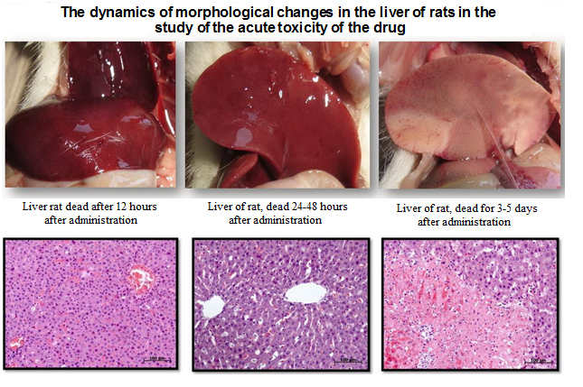
The introduction of large volumes and high doses of drugs in the acute toxicity studies are often accompanied by dystrophic and necrotic lesions of liver tissues, which can be observed in postmortem histological examination of cadaveric material.
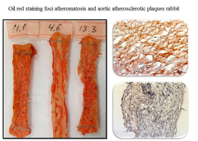
In the study of atherosclerosis applied macroscopic and microscopic staining of the aortic intima dye Oil Red (oil red), which allows good visualization of lesions atheromatosis.
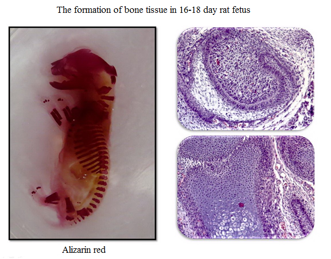
In experiments to study the reproductive and fetal toxicity deviations in osteogistogeneze fetuses to determine who carried out staining of cartilage and bone skeletons Alizarin red.
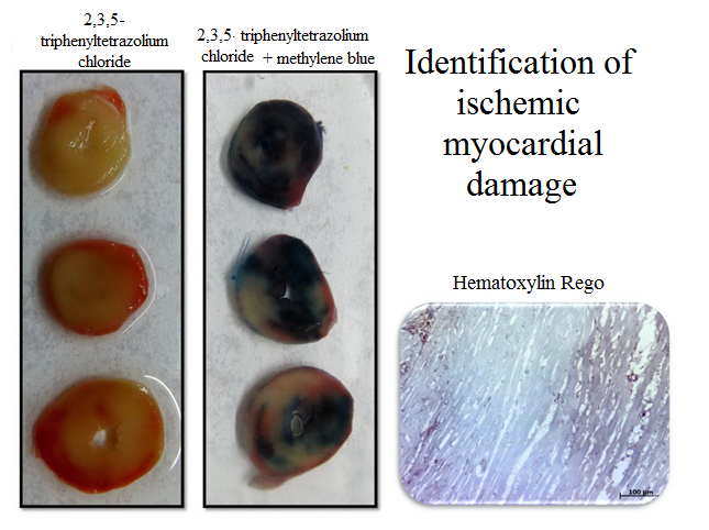
To determine myocardial ischemic lesions we use complex macroscopic and microscopic methods coloring material and the coloring 2,3,5-triphenyltetrazolium chloride based on determining the overall activity of the tissue dehydrogenase.
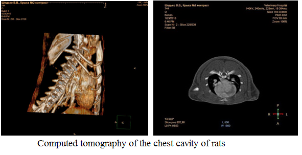
Computed tomography (CT) of organs and tissues of the experimental animals is one of the most promising areas of instrumental diagnostics both in terms of a more complete picture of the disease, and from the standpoint of the humane treatment of animals. CT allows assessment of his lifetime lesions, study the dynamics of the disease and the effectiveness of treatment without the need evtanazirovaniya animals at all interested control points of the experiment. At the same time, the use of CT significantly expands the notion of localization of various lesions, the degree of their severity and pathogenesis of the disease, allows to obtain reliable data, correlated with the results of conventional morphological studies in real-time.
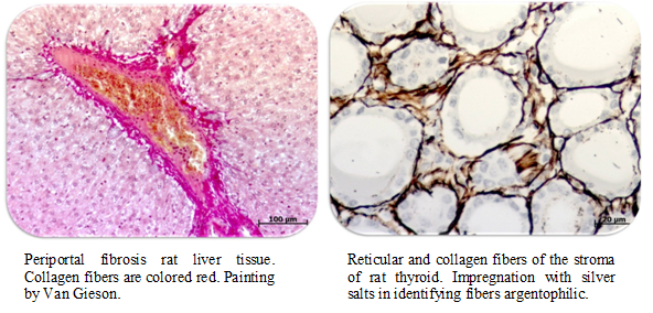
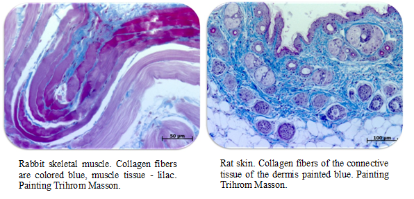
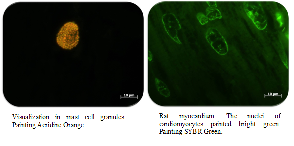
Histochemical methods of research for many years are used by researchers in the study of the normal structure of organs and diagnosis of pathological states. Histochemical staining techniques can detect tissue structures of interest, to identify and evaluate morphometrically organ damage such as tissue fibrosis, muscular dystrophy, and other pigments deposits. However, they do not exhibit species specificity, which is a great advantage when for different types of animal studies.
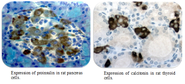
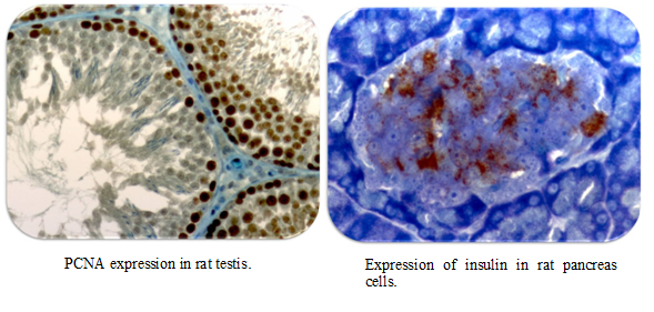
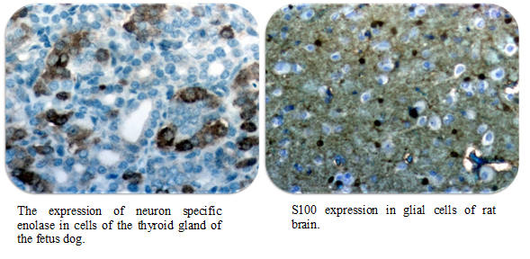
Immunohistochemistry studies of organs and tissues of animals used for experimental and diagnostic solutions tasks received in recent years widespread mainly due to the selectivity and high sensitivity of the used markers. These methods are relevant and most informative in studies on diabetes, neuropathy, pathologies of the cardiovascular system and endocrine organs.
In our work we use a wide range of immunohistochemical markers that are designed to not only the architecture of the tissue structure of the cells, but the mechanism of action study drugs.
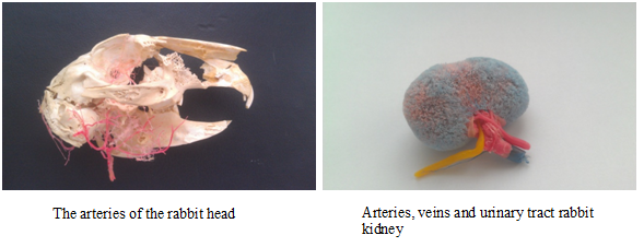
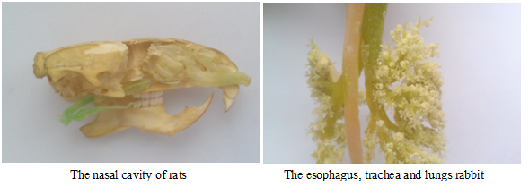
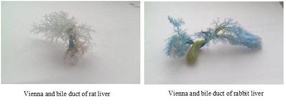
The most important tasks of anatomical science and morphology as a whole is to define interspecies differences in the structure and syntopy organs in animals. These data are the basis for the development of surgical approaches and the implementation of a large number of experimental conditions such as ligation of arteries and veins, intravascular injection, and others. The information as relevant when selecting dosage forms that are often associated with the anatomical structure of organs in animals.
We aim to expand the existing knowledge of comparative anatomy and experimental animals receive the most striking anatomical specimens showing the structural features of the bodies of animals which can not be seen at autopsy.
The material is subjected to morphological studies, subject to archiving for 1 year in accordance with the Russian Ministry of Health Order from 06.06.2013 N354n “On the procedure for post-mortem autopsy.”
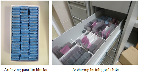
Histological archive contains all the necessary information about the experiments to allow retrospective analysis micropreparations, paraffin blocks and organs of animals.
To archive animal organs we use HistoSafe system. The material is placed in a strong plastic bag containing 10% buffered formalin, after which the package is sealed and organs throughout the backup period is stored in a sealed package.
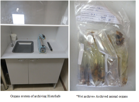
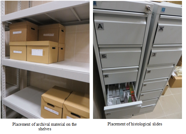
Histological slides and blocks are archived in separate shelves to store the identification numbers of the experiment.
Histological archive room is equipped with shelves, on which is placed the material to be stored.
-
Ustenko Zh.Yu., Loseva E.A., Savvateikina A.I. Determination of the decalcification time of sternum, knee joint and nasal cavity specimens from mice, gerbils and hamsters. Laboratory Animals for Science. 2023. №2. P. 66–73. https://doi.org/10.57034/2618723X-2023‑02‑06. Abstract. Procedure of decalcification necessary for histology of bone tissue. Decalcification is the procedure of removing minerals from the bone organic matrix. Solutions of varying acids usually used as a decalcification solution. Whole bone or piece that had been cut out preliminary are can be sampled for decalcification. Study of a knee joint and nasal cavity specimens usually uses in preclinical research. Also, for the study of bone marrow, a sternum often is sampled. The cutting out of small piece often leads to the destruction of the sample, because of their small size. Thus, whole maxilla, whole sternum and knee joint to include femur, fibula and tibia are more often sampled for research. This article describe a time of decalcification above-mentioned specimen that were sampled from mice, gerbils and hamsters. Solutions of formic and hydrochloric acids were used as the decalcification solution. The decalcification solution were changed every other day. The severity of decalcifications and specimen readiness for histology procedure were evaluated by gravimetric method. We found that the trends in mass change and decalcification time change were the same for all animal types noted above. In none case, the absolute severity of decalcification was reached, within 10 days. However, the severity of decalcification of whole specimens of bone was sufficient to make a quality histological slide. All specimens achieved the sufficient severity of decalcification on 4th day. This period allows to evaluate the morphology of the respiratory and olfactory epithelium of the nasal cavity, joint cartilage and bone marrow cells. The period of decalcification can be extended to 7 days, if anatomic and topographic features and them the best quality more important. The period of decalcification more 7 days leaded to irreversible change of cell morphology. Cells had poor detalization, cell nuclei were stained with hematoxiline badly, cell borders were unclear. [Full text is available in Russian].
-
Gushchin Ya.A., Makarova M.N., Shabanov P.D. Effect of intragastric administration on the morphology of laboratory rats’ gastrointestinal tract. Reviews on Clinical Pharmacology and Drug Therapy. 2023. 21(1). P. 57–68. DOI: https://doi.org/10.17816/RCF21157-68
-
Rekomendatsii po provedeniyu nekropsii laboratornykh zhivotnykh: monografiya / Pod red. V.G. Makarova i M.N. Makarovoi. — Sankt-Peterburg: NPO «DOM FARMATsII», 2023. — 92 str. p ill. [Full text is available in Russian].
-
Gushchin Ya.A., Shabanov P.D., Gushchina S.V. Pathomorphological changes in the gastrointestinal tract of Wistar rats with repeated intragastric administration of vehicles used in preclinical studies. Laboratory Animals for Science. 2022. №4. P. 10–20. https://doi.org/10.57034/2618723X-2022‑04‑02. Abstract. Оne of the most important tasks facing the developer of new medicines is not only to assess the activity of a potential medicinal substance, but also to test for safety, including its finished dosage form (FDF). Taking into account that the oral administration is the most common route, it is also widely in demand in preclinical studies. However, undisturbed tablets or capsules are not suitable for many types of animals, especially small mice and rats. Therefore, solid FDFs in most cases have to be converted into liquid forms. For this purpose, a wide range of different vehicles are used water, vegetable oil, starch gel, syrups, as well as surfactants, such as TWIN. Often these vehicles have to be used in large volumes, so they should be well tolerated by animals and not cause side effects. However, in our work, we sometimes find pathologies of organs and tissues in control animals that cannot be explained by the effects of the tested substances or models, background pathology. In literary sources, especially in Russian, there is very little precise data on the effect of vehicles on the organs of the animals gastrointestinal tract, and the authors limit themselves to conclusions: “non-toxic” or “well tolerated”. In this regard, we were tasked with identifying and determining the prevalence of background diseases, as well as the frequency of pathomorphological changes in the gastrointestinal organs of Wistar rats in response to intragastric administration of vehicles most often used in our research center. A retrospective analysis of preclinical studies was carried out, such vehicles were investigated as water, 1% starch gel, 0.5% methylcellulose, 64% sugar syrup, corn oil and 1% TWIN-80. As a result, it was revealed that the procedure of intragastric administration directly has a traumatic effect in the upper gastrointestinal tract. At the same time, starch gel and methylcellulose contribute to damage to the mucous membrane of the esophagus with the subsequent development of tissue inflammation. Corn oil, sugar syrup and TWIN, on the contrary, facilitate the introduction, but in the lower gastrointestinal tract provoke enteropathy, as well as metabolic disorders of the liver. The obtained data will subsequently help to differentiate the background pathology from the pathology caused by exposure to the tested substances, which will significantly improve the detection of possible undesirable toxic effects of drugs. [Full text is available in Russian].
- Ustenko Zh., Petlitskaya S., Gushchina S., Gushchin Ya., Makarova M. The effect of fi xing solutions on the severity of artifacts in the retina and choroid in histological sections of the rabbit eye. Laboratory Animals for Science. 2022. №1. P. 26–33. https://doi.org/10.29296/2618723X-2022-01-04 Abstract. Rabbits are often chosen as animal models for the study of eye pathologies, including pathologies of the retina and choroid. Fixation of the eyeball of a rabbit is a diffi cult task. In the process of fi xation, many artifacts occur in it. Many factors influence the frequency of occurrence and severity of artifacts: the procedure for selecting the eyeball, the type of fi xative fluid, and the cutting method. The most common artifacts in the study of histological sections of the eyes of rabbits include separation of neuroretina from the pigment epithelium, separation in the choroid, separation between the choroid and sclera, bleb-like protrusions of the nerve fi ber layer into the vitreous. The current study presents the result of the evaluation the effect of the type of fi xation fluid on the severity of the main artifacts in the retina and choroid of the eyes of rabbits. We studied the effect of 4 types of fi xing fluid: 10% neutral formalin, Davidson’s solution, freshly prepared 4% paraformaldehyde solution, freshly prepared 2% paraformaldehyde solution. Retinal separation was noted in all samples on macroscopic examination. Macroscopically the greatest severity of the separation was detected in samples fi xed in 10% formalin solution. Histological examination showed that samples fi xed in freshly prepared 2% paraformaldehyde solution had greatest severity of the separation of neuroretin from the pigment epithelium (p=0.0009). Samples fi xed in 10% neutral formalin solution had greatest severity of the separation of the choroid (p=0.0004). There was no statistically signifi cant effect of the type of fi xative fluid on the severity of separation of the choroid from the sclera (p=0.0750). Bleb-like protrusions of the nerve fi ber layer was found in groups fi xed in freshly prepared 4% paraformaldehyde solution, 10% formalin solution and Davidson’s solution once. The statistically signifi cant was found between the osmolality of the fi xing solution and the severity of retinal dissection in histological sections. Spearman correlation coeffi cient – 0.556 (p0.05). [Full text is available in Russian].
- Gushchin Ya.A., Belyaeva E.V., Ustenko Zh.Yu. Dynamics of post-mortem changes in the bodies of dead laboratory animals. Laboratory animals for scientific research. 2021; 4:69–81. https://doi.org/10.29296/2618723X-2021-04-08 Abstract. Patho-morphological study of organs and tissues it is an important tool in modern biomedical research. It is considered an integral part of the development, testing and safety assessment of new medicinal substances. However, due to various reasons, it is not always possible to perform an autopsy immediately, and there is a necessity to preserve the corpses for further study. The purpose of the study was to evaluate the dynamics of postmortem changes in laboratory animal’s corpses (mice, rats and rabbits), to identify optimal conditions for the storage (+16–22, +2–4 or -18–22°C), and to determine the timeframe for postponing necropsy without loss of informativeness (from 12 to 72 hours and 2 months
in case of freezing). Post-mortem changes were analyzed macroscopically and microscopically using a semi-quantitative assessment. It has been found that changes occur faster when corpses stored at room temperature. Besides, the size of animals also affects on the rate of decomposition. So in mice, with their small body volume, autolysis processes begin to develop in 18 hours after death, and by 72 hours they become catastrophic. At the same time, in larger animals – rats and rabbits, the development of post-mortem changes is slightly delayed, so after three days stored at a temperature of +16–22°C, macroscopic diagnosis was still possible. The decomposition processes can be signifi cantly slowed down by placing the corpses in a refrigerating chamber with a temperature of +2–4°C as soon as possible. But although for macroscopic
analysis, the corpses of all the studied animal species are preserved two and, albeit somewhat worse, three days after death, the sampling of tissues for histological examination should be carried out within 48 hours from the moment of death. Storage of corpses at a temperature of -18–22°C is an extreme measure, because, although the macroscopic structure of organs does not change in any way, the microstructure of tissues is disrupted, due to cryogenic damage. Thus, as a result of the study, the optimal time for the possible pathomorphological examination of the laboratory animals corpses after their unplanned death without loss or with minimal loss of informativeness under various storage
conditions was revealed. [Full text is available in Russian]. - Ustenko J.Yu., Vikulina D.A., Gushchin Ya.A. Determination of the decalcifi cation time of rat sternum, knee joint and nasal cavity specimens. Laboratory Animals for Science. 2021. №3. P. 17–26. https://doi.org/10. 29296/2618723X-2021-03-03 Abstract. Вone tissue and calcifi ed tissue need preliminary decalcifi cation before histological processing. Decalcifi cation is the technique for removing minerals from prefi xed tissue samples. Different solutions are used for decalcifi cation depending on the type of study, the time allotted to work and the type of sample. It is important to determine the “end point” of decalcifi cation. The end point is the moment when all inorganic material is removed from the tissue, leaving the remainder of the organic matrix intact. There are a number of methods for determin ing the end point. For example, manipulation (probing and bending), weighing, radiography. In hospitals end point control is usually performed for each batch of samples. In preclinical studies, there are many samples containing bone tissue, they are a routine practice, the procedure for their selection is standardized within a specifi c laboratory, as a rule. In preclinical studies, there are many samples containing bone tissue, they are a routine practice, the procedure for their selection is standardized within a specifi c laboratory as a rule. These features allow standardizing the decalcifi cation method and determining control points for typical samples using one of the methods listed above. This article presents a comparative characteristic of methods for determining the end point of decalcifi cation and establishes the decalcifi cation time of rat sternum, knee joint and nasal cavity specimens. The weighing is most suitable method for monitoring end point in routine preclinical studies. It is accurate, simple to perform, fast, and available to most laboratories. The optimal decalcifi cation period for samples of the sternum, knee joint and nasal cavity – 6 days – was established when using a solution based on hydrochloric and formic acids and in accordance with the described sampling procedure – the optimal decalcifi cation period for samples of the sternum, knee joint and nasal cavity was established when using a solution based on hydrochloric and formic acids and in accordance with the described sampling procedure. The shorter time of the samples in the decalcifying solution results in thick sections with more artifacts. A longer time leads to the loss of normal cell morphology and their ability to be stain. It is necessary to validate the decalcifi cation method in the laboratory, taking into account the reagents, the type of samples and the method of their selection. [Full text is available in Russian].
- Gushchin Y.A. Сomparative anatomy of the experimental animals and human heart. Laboratory Animals for Science. 2021. №1. P. 56–67. https://doi.org/10.29296/2618723X-2021-01-06 Abstract. The problem of cardiovascular system diseases treatment and prevention is still relevant. Pharmacotherapy plays an important role in the therapy, and, consequently, the development of new drugs. One of the invariable development stage is preclinical research involving laboratory animals determining effectiveness and safety of new drugs. In this paper we tried to put together the basic elements of human and laboratory animals heart structure, and to compare heart morphology to determine similarities and differences in the anatomy and histology of humans and different animals species. Anatomy of the mammalian heart has the same structural plan. Topographic location and anatomical and histological structure of the organ are similar. However, there are also some signifi cant species features. Heart location is mediated by body spatial position. In tetrapods it shifts to the sternum and left ventricle turned to the anterior chest, in humans, on the contrary, the top of the heart is attached to the diaphragm, and the right half is turned anteriorly Comparing the anatomical structure of humans and laboratory animals heart, we can note a different number of certain anatomical structures, such as the valves, chords and papillary muscles. but this variety can also be traced within the species. The number and location of the hollow and pulmonary veins have pronounced features. The course of the coronary arteries and their contribution to the blood supply of the myocardium parts also has its own species specifi city. As a result the blood supply type of the myocardium is also different. The left coronary artery dominates, but in rats and mice right and left coronary arteries evenly supply blood to the myocardium. In addition, animals have separate septum branches of the right coronary artery, which are not present in humans. Also, in rodents, the coronary arteries almost immediately branches off the aorta and run into the myocardium thickness, in humans they run epicardially for a long time. The histological structure of the heart is the same in all species. There are small differences, rather related to the size of the animals. The conducting system of the heart in all mammals has a single plan of structure. And the species specifi city is manifested in the variability of the location of nodes and their cellular composition. In this review, only the morphological structure is considered and physiological parameters are not discussed, since this topic requires a separate review. [Full text is available in Russian].
- Ustenko Zh., Gushchin Ya. Methods of visualization of rat lymphatic vessels and nodes. Laboratory Animals for Science. – 2020. №4. P. 1-9 https://doi.org/10.29296/2618723X-2020-04-06 Тhe lymphatic system of animals and humans consists of lymph, lymphatic capillaries, vessels, ducts and lymphatic organs. It drainage and detoxification functions, and plays an important role in the formation of the immune response and metastasis processes. In the literature, the structure and topography of the lymph nodes are comprehensively described, but little attention is paid to the lymphatic pathways. The study of the lymphatic system is difficult on animal-biological models. Some of its components are difficult to identify. The lymphatic vessels are difficult to detect on macroscopic examination, even in large animals, since the lymphatic vessel wall is thin and the lymph is colorless. The lymph nodes in small rodents are difficult to study because of its small size. In addition, the lymph node tissue may not contrast well with the surrounding fatty tissue. The combination of these factors can lead to inaccuracies during the sampling and cutting of material. This article presents techniques for visualizing lymphatic vessels and lymph nodes in rats. We visualized the lymphatic vessels by subcutaneous injections of hydrogen peroxide, tinted with ink or acrylic blue paint. There was observed coloration of lymphatic vessels and regional lymph nodes in both cases. We injected the dye solution to animals immediately after death, after 2, 4, 12 and 24 hours. There was observed coloration of lymphatic vessels and regional lymph nodes in all corpses that’s been in storage for 2-24 hours, and only in one fresh corpse. The best visualization of the lymphatic vessels by this method was achieved in the limbs and tail. We were unable to reach reproducible results in the head, neck and trunk with this technique. We sampled for histological analysis lymphatic vessels painted ink. There was observed the coloration of the epithelium of the lymphatic vessel in one case. We visualized the lymph nodes by fixation in Davidson’s solution for 2 and 19 hours. The use of this method provided better visualization of the lymph nodes during cutting compared with visualization after 10% formalin fixation. In addition, fixation in Davidson’s solution has provided better detailing of the lymphoid cell nuclei [Full text is available in Russian].
- Tyutina K.V., Gushchin Y.A., Makarova M.N., Makarov V.G. Evaluation of bone marrow in preclinical studies // Translyatsionnaya meditsina=Translational Medicine. 2020. Vol. 7 (5). P. 119-130 DOI: 10.18705/2311-4495-2020-7-5-119-130 Background. The study of red bone marrow in preclinical studies is an important component of the study program of the drug. Objective. To compare the results obtained in parallel studies of the material from the same animal by two different methods for rats, rabbits and mice (cytological examination of the material obtained from the femur and histological examination of the material obtained from the sternum). Design and methods. Bone marrow obtained from intact animals was studied. Cytological preparation was stained according to Romanowski–Giemsa, histological preparation was prepared in the usual way and stained with hematoxylin-eosin. Stained preparations were examined by microscope. Results. As a result of this study was sets of myelograms with leuko-erythroblast ratio and neutrophil maturation index. Conclusion. A number of conclusions were made: when conducting preclinical studies, the most appropriate can be considered a bone marrow sampling from the femur; the fence can be carried out both for cytological examination, and for histological; taking into account the high variability of indicators in preclinical studies of myelograms, it is necessary to have control groups of (intact) animals of the same species that did not receive the study drug; Myelograms in rabbits turned out to be the least varied, both according to the results of our own studies (with histological and cytological studies), and when compared with literature data, which allows us to recognize rabbits as the best object for studying the composition and function of red bone marrow; when comparing the literature data, significant patterns in the number of cells at different age periods were not observed in the studied animal species, and, therefore, in preclinical studies using KKM analysis, animals of any age can be used [Full text is available in Russian].
- Belyaeva E.V., Guschin Ya.A. The methods of visualization and research of the git-associated lymphoid tissue of laboratory animals. Laboratory Animals for Science. 2020. №3. https://doi.org/10.29296/2618723X-2020-03-09 The immune system is an important part of the vital activity in the organism, because it prevents a penetration of foreign bodies and substances or it deletes them very quickly. Its elements can be found throughout in all organ systems in the form of various lymphoid formations or individual cells. The mucosa-associated lymphoid tissue (MALT) is one of the important parts of the immune system. The gut-associated lymphoid tissue (GALT) is the largest department of MALT, because about 80% of all defensive cells contact with the mucous membrane of the intestine. Some toxic effects on the organism show expression or suppression of immune responses of a body or don’t appear. So thorough lymphoid tissue analysis is a very important part in the preclinical study. The purpose of this study is to detect of existing visualization methods and research of the GALT and a selection of the most optimal methods for their usage in experiments. Some macroscopic research methods were considered, one of which was put into practice. The organs of male and female rats and rabbits were used in the study. A detailed description of visualization methods has not been preserved in literary sources, so the most optimal method to stain native intestinal preparations for macroscopic examination of intestinal associated lymphoid tissue was developed in experiments. The most informative method of the histological examination also was described and shown in this research, which one allows to study and the GALT and the intestinal mucosa. In this case, you need to select all the sections of the intestine, so that can be explored on all surface of the intestinal mucosa, and you can also examine in detail all the lymphoid formations of the intestine under a microscope. The results will help to expand the methods of visualisation and researching of the immunotoxicity of various drugs in preclinical studies. In addition, the results can be used in a detailed study of the structure of the immune system of an animal’s organism [Full text is available in Russian].
- Gushchin Y.A., Kryshen A.A.Testing of an infective inflammation model of the gastrointestinal tract, associated with helicobacter pylori, in laboratory gerbils. Laboratory Animals for Science. – 2020, №3. https://doi.org/10.29296/2618723X-2020-03-08 Peptic ulcer of the stomach and duodenum is a chronic recurrent disease that occurs with alternating periods of recrudescence and remission. The main manifestation of this disease is the formation of a defect in the wall of the stomach and duodenum. One of the main factors in the occurrence of peptic ulcer disease is infection with Helicobacter pylori (H. pylori). These are microaerophilic, non-spore-forming, gram-negative, curved rod-shaped or coccoid bacteria. They play an important role in enhancing the aggressive properties of gastric contents and weakening of gastric and duodenal mucosal defense. The high frequency of chronic Helicobacter gastritis causes a high incidence of cancer. Therefore, the development of a model of Helicobacter-induced gastroduodenal diseases in vivoto search for alternative therapy for H. pylori infection is currently relevant. Thus, the aim of the study was to develop a model of infectious (associated Helicobacter pylori) inflammation of the gastrointestinal tract. Assessment of the developed pathology was carried out by determining microscopic changes in the tissues of the gastrointestinal tract. 30 Mature male laboratory gerbils were used as a test system. Positive control animals were infected with a suspension of fresh H. pylori culture at a concentration of 2×10⁹ CFU / ml in a volume of 0.5 ml with intragastric administration twice, once a day, for 2 days. Sterile trypton soy broth (a medium for H. pylori cultivation) was gavaged to negative control animals according to a similar scheme. The morphological analysis was based on the international classification of chronic gastritis (the Sydney system and its Houston modification). As a result of the study, laboratory gerbils had a pathology of the gastric mucosa by the 24th week of the experiment. After 8 weeks, the infected animals registered initial manifestations of catarrhal gastritis. By the end of the 16th week of the experiment, in addition to increasing gastritis, bacteria corresponding to H. pylori in their morphological forms were identified. By the end of the study, there was an inflammatory component in the gastric mucosa, atrophy of the glands, and erosive and ulcerative lesions of the mucous membrane. Bacterial forms corresponding to H. pylori was identified with a specific staining in the mucous membrane of the stomach. Pathological changes were observed mainly in the antral part of the stomach. The chosen system for assessing the severity of the pathology proved to be sufficient and objective, allowing us to fully analyze the development of pathology. However, the use of this strain of H. pylori did not lead to the formation of ulcerative and metaplastic changes when observed at 24 weeks of pathology development, which was expected based on the literature data. Also, during this period of time, it was not possible to achieve the development of duodenite. It is likely that the used strain of bacteria did not have sufficient virulence, which should be taken into account in future studies [Full text is available in Russian].
- Ustenko J.U., Gushchin Y.A. A review of the most common pathologies of the stomach in laboratory animals in preclinical studies of drugs. Laboratory Animals for Science. – 2020, №2. https://doi.org/10.29296/2618723X-2020-02-02 Pathomorphological analysis is an integral part of preclinical studies. It allows evaluating the pathological changes that occur in the organs of laboratory animals at macroscopic and microscopic levels. Gastric pathology can develop due to the local irritating effect of the test articles during peroral and intragastric administration, as a spontaneous or background pathology, due to various mechanisms with other routes of administration (intramuscular, subcutaneous, intraperitoneal and others). The classification and morphological criteria of gastric pathology in laboratory animals are most fully developed by the INHAD group for rats and mice. Clear recommendations have not been developed for other animal species yet. Orientation of toxic pathologists to a unified classification will be avoiding confusion in interpretations, will be ensuring reproducibility of results, and will allow comparing of the results of various researchers. А possible classification of gastric pathologies in laboratory animals in preclinical studies is presented in the article. It is based on an analysis of literary sources. Congenital pathologies such as squamous cysts and ectopic tissues are described. Atrophic and hypertrophic changes in the mucous membrane; metaplastic changes; dystrophic changes, such as vacuolization; detection of eosinophilic globules, mineralization, amyloidosis; the formation of glandular cysts and diverticulum in the epithelium of the stomach are considered. Аpplication of additional microscopic examination techniques described: immunohistochemical staining for TFF2, CDX1 and CDX2 for differentiation of metaplastic shifts; Congo red stained to verify the diagnosis of amyloidosis. Apoptosis and necrosis of а stomach epithelium are also described in detail. Since these processes are indistinguishable during the evaluating routine sections stained with hematoxylin-eosin, additional methods (transmission electron microscopy, DNA-laddering, TUNEL, immunohistochemistry for caspases, (in particular, caspase-3) are listed. They allow to accurately differentiate apoptosis and necrosis. Inflammatory changes are described: inflammatory infiltrate and inflammation. А gastric dilatation, volvulus, detection of trichobesoars in the lumen of the stomach also are considered. For each pathology, an illustration, a brief macroscopic and microscopic characteristic is given. Possible causes of stomach pathologies in rats, mice, hamsters, gerbils, ferrets, rabbits, dwarf pigs in preclinical studies are described[Full text is available in Russian].
- Gushchin Y.A., Kvanchiani V.V. Comparative morphology of the oral cavity in experimental animals and humans // Laboratory Animals for Science. 2020. № 1. https://doi.org/10.29296/2618723X-2020-01-02 In pre-clinical studies on laboratory animals the most often studied substances are oral. The data for different types of laboratory animals is very contradictory and ambiguous. However, the differences between them can have a significant effect on the obtained result. Analysis of the literature data revealed the main features of anatomically and histologically similarity of the human’s and laboratory animals’ oral cavity structure. The oral cavity is the initial section of the digestive system, where the food lump undergoes mechanical and chemical treatment. Development of the face and mouth is carried out on the same principle of common embryonic rudiments. Anatomically and histologically the oral cavity of the examined species has similar structures. But the structure of the oral cavity was greatly influenced by the nature of the familiar food. First of all the most differences can be noted in the structure of the teeth and the dental formula. So, for example, in humans and rabbits, there are two consecutive generations of teeth, while the remaining animals have only one set. In humans, rodents and hares different and the structure of molars. A cardinal difference was found in the histological structure of the oral mucosa. In humans and partly in rabbits the lining represented by neorogovevayuschy multilayered epithelium, whereas in rodents it keratinized in all departments, also varies the thickness and number of layers. by a multilayered non keratinized epithelium. While in rodents it is keratinizing in all departments and the thickness and number of layers are differ. The blood supply, lymph drainage and innervation of the oral cavity occur in the same way in humans and animals. This comparative review will help in the planning of studies and further work with the obtained data, and will also be useful for histologists, physiologists and pathologists working with laboratory animals [Full text is available in Russian].
- Muzhikyan A.A., Shed’ko V.V., Zaikin K.O., Gushchin Ya.A. , Makarova M.N., Makarov V.G. Comparative morphology of salivary glands of humans and laboratory animals // Morfologiya. 2020. Vol. 157. № 1. P. 79-92. doi: 10.34922/AE.2020.157.1.013. The article summarizes comparative data on the normal morphology of the parotid, submandibular, and sublingual salivary glands of humans and some laboratory animals most commonly used in biomedical research. The information presented in this review shows the similarity of the general principles of the structure and function of the large salivary glands of humans, rats, mice, rabbits, guinea pigs and hamsters. However, morphology is still quite variable, which is explained, first of all, by phylogenetic features associated with the lifestyle of humans and animals, as well as the type of nutrition. When the anatomy of the glands is principally the same, only slightly different in shape and relative size, the interspecies differences in the topographic location of the glands and their microscopic structure became more significant. The differences in the composition of the secretion of human glands and glands of studied animals were mainly due to the structural features of the acini and excretory ducts and the location of myoepithelial cells in these structures. Thus, in preclinical studies of drugs, it is necessary to take into account not only the physiological characteristics of salivation and the biochemical composition of saliva but also the structural features of the salivary glands of various animal species. The information on the anatomy, topography, and syntopy of the salivary glands presented in this review can be useful in performing surgical procedures, in diagnostic and treatment procedures, and for modeling pathologies. Information about the histological structure will help to avoid difficulties in making a diagnosis, as well as incorrect interpretation of the data [Full text is available in Russian].
- Belyaeva E.V., Ustenko J.U., Guschin Ya.A. Technique of dissection and extracting organs of laboratory animals. Message 6 – minipigs. Laboratory Animals for Science. 2019. №4. P. 55-77 https://doi.org/10.29296/2618723X-2019-04-08 The sixth report from a series of articles on the methodology of necropsy and organ extraction o laboratory animals is presented. The report describes and illustrates in detail the methodology for sequential and complete necropsy and organ extraction of miniature pigs. Currently, miniature pigs are widely used in biomedical research as laboratory animals, since their anatomical and physiological characteristics are most similar to human ones. Miniature pigs are considered among the basic models for studying stroke, diabetes, organ transplantation, surgical treatment methods, and also used for testing of pharmaceutical products and biomechanical devices. Also, these animals are essential in studies of the structure and physiology of skin, cardiovascular, digestive and urinary systems. The size and weight of minipigs are perfect for modeling of many pathologies. Also they provide a sufficient amount of biomaterial, e.g. nternal organs, blood or other body fluids, for further laboratory researches. This article describes the step-by-step procedure of animal necropsy from preparation, external examination, sampling of mammary glands and skin, sequential opening and examination of the thoracic and abdominal cavities, and to the extraction of separate systems and organs. Possible methods of extracting organs of the oral cavity, thoracic, abdominal and pelvic cavities are described and illustrated, and the fastest, most effective and safe method of extraction of the brain from the cranial cavity is presented. The methods of eye extraction along with the optic nerve and the extraction of muscles and adjacent peripheral nerves are demonstrated. All methods of organ extraction, as well as the sequence of actions described in this article minimize damage of harvested organs and prevent their contamination in order to avoid the formation of some artifacts, which might be found during subsequent histological examination and could lead to conflicting results [Full text is available in Russian].
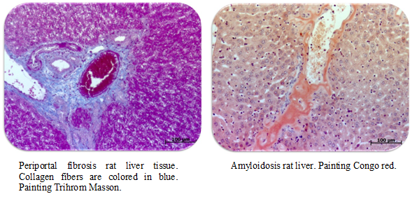
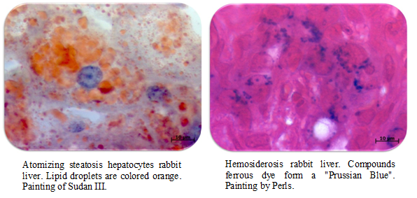
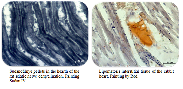
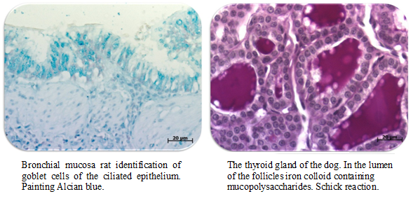
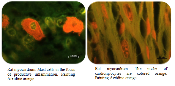
- Gushchin Y.A. Additional histological methods of stainig in preclinical studies // Laboratory Animals for Science. 2019. 4. P. 44-54 https://doi.org/10.29296/2618723X-2019-04-07 In clinical practice and in pharmacological studies, microscopic analysis is an integral part of the study of normal structure of tissues and pathologically altered organs. To prepare the material for histological examination, visualization is carried out by staining the tissue with dyes. The most common routine staining is hematoxylin and eosin, which is not enough to reveal the full picture of the process. Therefore, it is necessary to use a number of additional stains. Some of them can be used as routine instead of classical hematoxylin and eosin, for example, van Gieson stains or Mallory trichrome. Most stains are more specific, and they are used to identify specific structures or chemical compounds in cells and tissues. This allows you to obtain a significant amount of information, which facilitates an understanding the course of normal and pathological processes. Methods for the detection of mucopolysaccharides with Alcian blue are used in the study of the gastrointestinal tract and respiratory system. PAS staining is necessary in the diagnosis of accumulation diseases, some oncological processes and fungal infections. Fat-soluble dyes, primarily Sudan III and scharlach red are used in the study of dystrophic diseases and atherosclerosis. The Oil Red O dye is applicable for macroscopic assessment of the area of atherosclerotic lesions of the aorta. Congo red is the most popular dye for the amyloid detection. Specialized dyes for the diagnosis of myocardial damage were considered, such as (HBFP) (Haematoxylin-Basic Fuchsin-picric acid) and tetrasolium salts to visualize the area of damage to the heart muscle and brain. In this review, we examined several histological dyes, some features of their application, as well as mechanisms of the dye-substrate interaction in tissues. These staining methods can be used during histological work, and consider the possibility of their use, which will help to identify, as well as comprehensively study the pathological processes modeled in preclinical studies [Full text is available in Russian].
- Koptyaeva K., Ustenko J.U., Belyaeva E.,Guschin Ya.A., Makarova M., Makarov V. Technique of dissection and extracting organs of laboratory animals. Message 5 – rabbit, ferret // Laboratory Animals for Science. 2019. №3. https://doi.org/10.29296/2618723X-2019-03-05 The fifth message from a series of articles on the methodology of necropsy and extraction of organs of laboratory animals, describing in detail and illustrating the technique of sequential and complete necropsy and extraction of organs of laboratory rabbits and ferrets, is presented. Rabbits are suitable for long-term ongoing research, and are the only laboratory rodents from which you can get recombinant pharmaceutical proteins and explore the oral or intragastrically forms without destroying the integrity of the tablets. In addition, they are used to develop and study new surgical methods, physiological tests and studies of various types of toxicity for drug testing. Various viral and bacterial diseases were studied on ferrets, as well as pathology of the organs of hearing, vision, the vomiting reflex due to the similarity of many anatomical, metabolic and physiological characteristics to humans. This article describes the method of necropsy from the procedure of external examination of a corpse, sequential dissection and examination of the thoracic and abdominal cavities, and before the extraction of organs, the selection of various bone preparations. Possible methods for the extraction of organs of the oral cavity (including extraction of the tongue, pharynx), all organs of the thoracic, abdominal and pelvic cavities are described and illustrated. Describes the method of extraction of the brain, the method of selection of the chest bone for histological examination of the bone marrow and upper jaw for histological examination of the nasal passages. A procedure for preparing a spinal cord site for fixation in formalin without removing it from the spinal canal is described. Demonstrated methods for extracting the eyes, together with the lacrimal gland and eyelids by a single organocomplex, extracting the muscles and adjacent peripheral nerves. The methods of extraction and the sequence of actions performed, described in this article, minimize the damage to the organs being removed and prevent their contamination in order to prevent the occurrence of certain artifacts detected by subsequent histological examination [Full text is available in Russian].
- Guschin Ya., Kovaleva M. Comparative Morphology of Human and Laboratory Animals. Laboratory Animals for Science. – 2019. №2. https://doi.org/10.29296/2618723X-2019-02-06 Knowledge the characteristics of the morphological structure, the main structural and functional components of human skin and laboratory animals is the foundation of rational planning of pharmacological and toxicological preclinical studies of skin dosage forms in aspect. The choice of laboratory animals must be justified for accurate translation of the results of pre-clinical studies into clinical studies (early phase). A brief report shows the comparative morphology of human skin and certain types of laboratory animals: mouse, rat, guinea pig, hamster, rabbit, ferret, minipig. Structural similarities and differences of skin layers (epidermis, dermis, gmpoderm) are described. A comparative characteristic of the thickness of the epidermis and dermis in different species, carried out on the analysis of literature data and results obtained in their own research. A comparative analysis was conducted both between humans and laboratory animals, and between species.[Full text is available in Russian].
- Koptyaeva K., Guschin Ya., Belyaeva E., Makarova M., Makarov V. Technique of Dissection and Extracting Organs of Laboratory Animals. Message 4: Guinea Pig, Gerbil, Degu. Laboratory Animals for Science. – 2019, 2. https://doi.org/10.29296/2618723X-2019-02-05 The fourth message from the series of articles on the methodology of necropsy and extraction organs of laboratory animals, describing in detail and illustrating the technique of sequential and complete autopsy and extraction of organs of laboratory guinea pigs, gerbils and degu. Guinea pigs are often chosen to study the allergenic properties of drugs and to determine the pharmacological activity, for example, in experiments that model hypercholesterolemia, airway hyperresponsiveness, etc. The high susceptibility of gerbils makes it possible to be a model for various infectious diseases. Also on them examine the effect of drugs on the level of steroid hormones and cholesterol. Degus that predisposed to the development of diabetes mellitus are a good model for studying this disease. In addition to this, on them conduct studies of cataracts, atherosclerosis, and Alzheimer’s disease. This article demonstrates the procedure for necropsy of laboratory guinea pigs, gerbils, and degu. It describes the process of preparing an animal for an autopsy, conducting a primary examination of a corpse after euthanasia for the presence of external changes and injuries, fixing the corpse on the dissecting table, necropsy and examining the internal cavities of the body, as well as examining the superficial lymph nodes. Possible methods for the extraction of organs of the oral cavity (including extraction of the tongue, pharynx), all organs of the thoracic, abdominal and pelvic cavities are described and illustrated. The topographic location of some organs of the thoracic, abdominal and pelvic cavities is illustrated. Prescribed two ways to extract the brain, the method of separation of the chest bone for histological examination of the bone marrow and upper jaw for histological examination of the nasal passages. The procedure for preparing the spinal cord to formalin fixation without removing it from the spinal canal is described. Eye extraction with the lacrimal gland and eyelids, extraction of muscle and adjacent peripheral nerves is described. The methods of extraction and the sequence of actions performed, described in this article, minimize the damage of the organs being removed and prevent their contamination in order to prevent the occurrence of certain artifacts detected by subsequent histological examination. Conclusions are presented at the end of the article.[Full text is available in Russian].
- Vavilova V., Gushin Ya. Model of The Global Cerebral Ishemia in Mongolian Gerbil // Laboratory Animals for Science.– 2019. 2. https://doi.org/10.29296/2618723X-2019-02-03 ABSTRACT. The aim of this study was to develop a global cerebral ischemia simulation protocol (GCI, global cerebral ischemia) with subsequent reperfusion, which was carried out on Mongolian gerbil. The choice of a gerbil as a biological model is due to the anatomical feature of the blood supply to the brain. Gerbils, unlike other laboratory animals and humans, do not have a posterior communicating artery (do not have the full circle of Willis). Mortality, neurological status, pathological and histological studies were evaluated as criteria for the pathology degree estimation. Visualization of the ischemic region after the induction of pathology was carried out by staining brain sections with 2, 3, 5 triphenyl tetrazolium chloride (TTC). The method of the neurological status scoring which takes into account the presence or absence of pathological changes is described. The model is confirmed by pathological and histological examination. The severity of damage prevails on the 2nd and to a lesser extent on the 3rd day after the pathology induction. Histological examination of the gerbil brain identified neurons with signs of dystrophy (polymorphic, hyperchromic cells with fuzzy contours – “red” neurons) and reduced in size – “shadow cells”, as well as fields with mild gliosis in the cerebral cortex and cerebellar cortex. Thus, the results of the study showed that the 20-minute bilateral occlusion resulted in animal survival of about 60%, most animals demonstrated pronounced neurological deficit. Staining with TTC has shown that it is advisable to evaluate volume of the ischemic lesion in the first 24 hours after the induction of pathology. In addition, results showed that the neurological deficit was most pronounced during first 4 days after occlusion. To estimate long-lasting pathological changes it is advisable to conduct a histological examination of brain tissue samples [Full text is available in Russian].
- Koptyaeva K., Guschin Ya., Belyaeva E., Makarova M., Makarov V. Technique of dissection and extracting organs of laboratory animals. Message 3 − hamster // Laboratory Animals for Science. 2019. №1. P. 1-19. https://doi.org/10.29296/2618723X-2019-01-02 The third message from the series of articles on the methodology of necropsy and extraction organs of laboratory animals, describing in detail and illustrating the technique of sequential and complete autopsy and extraction of organs of laboratory hamsters. Among laboratory animals, hamsters occupy a special place due to the presence of large cheek pouches in their digestive tract. A large area of the mucous membrane lining the cheek pouches from the inside, allows to visually demonstrate and explore the local irritant effect of orally dispersible dosage forms (lozenges), which can easily be placed in the cavity of the cheek pouches. This article demonstrates the procedure for laboratory hamster necropsy. It describes the process of preparing an animal for an necropsy, conducting a primary examination of a corpse after euthanasia for the presence of external changes and injuries, fixing the corpse on the dissecting table, opening and examining the internal cavities of the body, as well as studying the surface lymph nodes. Methods for the extraction of organs of the oral cavity (including the extraction of the tongue, pharynx and cheek pouches along with the adjacent salivary glands), all organs of the thoracic, abdominal, and pelvic cavities are described and illustrated. The topographic location of some organs of the thoracic, abdominal and pelvic cavities is illustrated. Described two methods to extract the brain, along with olfactory bulbs, the method of separating the preparations of the chest bone for histological examination of the bone marrow and maxilla for histological examination of the nasal cavities. The procedure for preparing the spinal cord for fixation in formalin without removing it from the spinal canal is described. Described the extraction of the eyes together with the lacrimal gland and eyelids, the extraction of the muscles and the peripheral nerves adjacent to them. The methods of extraction and the sequence of actions performed, described in this article, minimize the damage to extracted organs, prevent their contamination in order to prevent the occurrence of certain artifacts detected during subsequent histological examination. Conclusions are presented at the end of the article [Full text is available in Russian].
- Koptyaeva K., Muzhikyan A., Guschin Ya., Belyaeva E., Makarova M., Makarov V. Technique of dissection and extracting organs of laboratory animals. Message 2 (mouse) // Laboratory Animals for Science. 2018. №4. P.1-24. https://doi.org/10.29296/2618723X-2018-04-05 Post-mortem examination of animals (necropsy, autopsy) is a diagnostic procedure aimed at finding out the cause of death of an animal, as well as determining the spectrum of macroscopically visible changes in organs and tissues of cadaver. In scientific and research practice, one of the key stages of the experiment and at the same time a humane experimental point often is euthanasia of animals with subsequent pathoanatomical and histological examination of organs. From the point of view of the quality and reliability the obtained data, an important step is the standardization of the procedure to dissection and extracting organs of laboratory animals. A series of articles about the carrying out of necropsy of laboratory animals will highlight the main stages of autopsy. In this work, a technique to dissection and extracting organs of mice is described [Full text is available in Russian].
- Zaikin K., Gayday E., Gayday D., Gushchin Ya., Muzhikyan A., Kryshen’ K., Makarova M. Spontaneous age-related pathology of the urinary system in rats // Laboratory Animals for Science. 2018. №4. P.1-15. https://doi.org/10.29296/2618723X-2018-04-03 The article is devoted to spontaneous age-related pathologies of the urinary system of rats detected in animals of control group in a long-term carcinogenesis experiment. Interest in this group of pathologies is relevant during long-term experiments since the elderly rodents’ death is often associated with oncological diseases and pathologies of the kidneys. Knowing of pathologies with spontaneous manifestation in laboratory rats is necessary for correct interpretation of experimental results as well as for the differential diagnosis of «spontaneous» and «induced» pathologies. For the classification of pathologies INHAND nomenclature was used. Histological examination of the organs of urinary system (kidney and bladder) was carried out in 200 males and 200 female Wistar rats aged 24 months. Tumor pathology in the article is not considered. Spontaneous age-related pathology of the urinary system in rats is very common – kidney pathology was found in all 200 males studied and in 165 of 200 females studied. Pathological changes considered as manifestations of chronic progressive nephropathy (such as interstitial inflammatory cell infiltration, glomerulosclerosis, basophilia and atrophy of renal tubule epithelium, tubular dilatation, cylinders in the lumen of tubules, focal interstitial sclerosis of the renal cortex) are most common; all 200 males studied and 140 of 200 females studied were identified. Vesicular dystrophy (vacuolization) of renal tubules epithelium is only pathology of urinary system that is more common in females than in males. Vesicular dystrophy has a diverse pathogenesis its clinical and biological significance is ambiguous it can precede necrosis of epithelium, but it can also occur in normal conditions. Pathology of bladder is less common it was detected in 22 males and 1 female. Among the pathological changes were protein plugs in the lumen of bladder and focal inflammatory cell infiltration of bladder. In the experiments described pathologies should be considered as spontaneous associated with age-related changes in the body [Full text is available in Russian].
- Gushichin Y., Myzhikyan A., Shedko V., Makarova M., Makarov V. Comparative anatomy of the upper gastrointestinal tract of experimental animals and humans // International bulletin of veterinary Medicine. – 2017, No. 3. – P. 116-129. Сomparative anatomy of the upper gastrointestinal tract of experimental animals and humans [Full text is available in Russian].
- Gushchin Y.A., Muzhikyan A.A., Selezneva A..I, Makarova M.N. Complex morphological evaluation of atherosclerotic damage of the aorta of rabbits in the experiment // Atherosclerosis and dyslipidemia. – 2017. 1. – P. 50-59.
- Muzhikyan AA, Khod’ko SV, Gushchin Ya.A., Makarova MN, Makarov V.G. Histological changes in the lymph nodes of rats during the modeling of acute cervical lymphadenitis // International bulletin of veterinary Medicine. – 2017, No. 1. – P. 75-83.
- Shedko VV, Gushchin JA, Muzhikyan AA, Makarova MN Retrospective analysis of histological archive // Development and registration of medicinal products. – 2016, № 3. – P. 162-165.
- Muzhikyan AA, Makarova MN The use of computed tomography in the evaluation of the state of organs and tissues of laboratory animals // InternationalBulletin of Veterinary – 2015, № 4. – P. 73-80.
- Katelnikova A.E., Kryshen K.L., Muzhikyan A.A., Makarova M.N., Makarov V.G. acute inflammation model: carrageenan air pouch // InternationalBulletin of Veterinary – 2015, №2. – P. 78-87.
- Kashkin V.A., Shekunova E.V, Muzhikyan A.A., Makarova M.N., Makarov V.G. Comprehensive assessment of the degree of pathology in modeling the adjuvant-induced arthritis in rats // InternationalBulletin of Veterinary – 2015, №1. – P. 92-103.
- Muzhikyan A.A., Makarova M.N., Gushchin J.A. Features histological working of organs and tissues of laboratory animals // InternationalBulletin of Veterinary – 2014, №2. – P. 103-108.
- Muzhikyan A.A., Makarova M.N., Gushchin J.A. Features mortem examination group of experimental animals // InternationalBulletin of Veterinary – 2014, №1. – P. 75-80.
- Khodko SV, Makarova MN, Makarov VG, Samusenko IA, Shirunova MG The experimental model of acute rhinosinusitis in rats to assess the therapeutic efficacy of drugs // Preventive and clinical medicine. – 2013. – T. 46, number 1. – P. 57-62.
- Proshin SN, Makarov VG, Makarova MN, Kryshen KL, Kovshin AV, Samusenko IA Ulcerative nonsteroidal anti-inflammatory drugs and hepatotoxicity of nimesulide in an experiment on rats // Surveys on the wedge. and lectures. therapy. –2012. – Vol.10, №1. –P.28-34.
- Tesakova SV, Samusenko IA, Karachinskaya IV, Kryshen KL, Abrashova TV, Makarova MN, Makarov VG The experimental model of cervical lymphadenitis in rats to evaluate the anti-inflammatory efficacy // Preventive and clinical medicine. – 2011. – №1 (38), pp 57-63.
- Tesakova SV, Makarova MN, Makarov VG, Samusenko IA, GushchinJA The effectiveness of the use of drugs with a high content of phytosterols in the treatment of chronic prostatitis // Bulletin of the Research Center of Volgograd. -2009. Number 2. C. 35-39.
- Makarova MN, Tesakova SV, Samusenko IA, Stolaschuk NV, Solovyov DE, Zhorina AS, Tikhonov VP, Makarov VG Experimental study of the effectiveness of biologically active food additives and drugs in a model of chronic prostatitis // Bulletin of St. Petersburg State Medical Academy. II Mechnikov. – 2007. № 2 (8). – P. 123-128.