Study of pharmacological agents affecting cardiovascular system
The study of hypotensive action of PA includes:
the assessment of PA influence on angiotension-converting enzyme (ACE) activity.
the study of antihypertensive influence of PA is carried out in spontaneously hypertensive rats (SHR) using the following models:
mezatonum model;
izadrinum model.
The study of PA efficiency in treating of coronary blood circulation disorders is performed in the following model:
the model of an experimental myocardial infarction.
The study of PA activity in pulmonary crotalin-induced hypertensia model;
The study of PA specific effect on blood vessels of chicken embryo chorioallantoic membrane includes an assessment of three mechanisms of PA action:
noradrenergic mechanism;
renin-angiotenzin mechanism,
endotelial mechanism (activation of endotelial NO-synthase).
Assessment of PA activity in diabetes model accompanied with an assessment of a capillary protective action is performed using the following models:
the model of an acute damage of the vascular bed by hydrogen peroxide;
the model of a prolonged diabetes complicated by micro- and (or) macroangiopathy;
Assessment of PA activity in the model of chronic heart failure (CHF) of a total type;
Assessment of PA activity in the model of CHF induced by sterile silicone oil (1 ml per 100 g body weight) administrated into both pleural cavities;
Assessment of cardioprotective effect of PA (cardiotonic and antiarrhythmic) in the isolated heart model by Langendorff Method;
Evaluation ofthe activity PA model dosed supravalvular stenosis in mini-pigs or rabbits;
Antiarrhythmic activity in the study of PA aconite, adrenalin, barium chloride or other models;
A study of antiarrhythmic activity of PA on models of ventricular arrhythmias;
A study of antiarrhythmic activity of PA on models of reperfusion arrhythmias;
Study antifibrillar activity of PA on mini-pigs, rats;
Assessment of hypolipidemic and antiatherogenic effects in the model of dislipoproteinemia.
The efficiency of test substances is estimated by the number of animals survived, ECG parameters and the blood pressure value, the biochemical indicators of peripheral blood, CSF and other tissues based on histology, histochemistry and immunohistochemistry analyses.
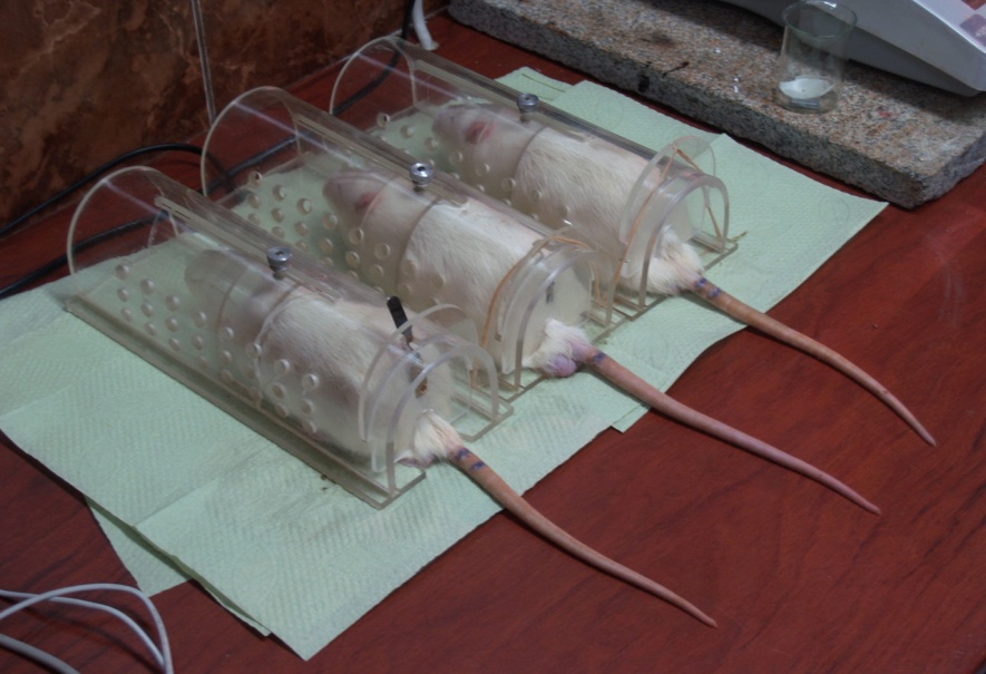
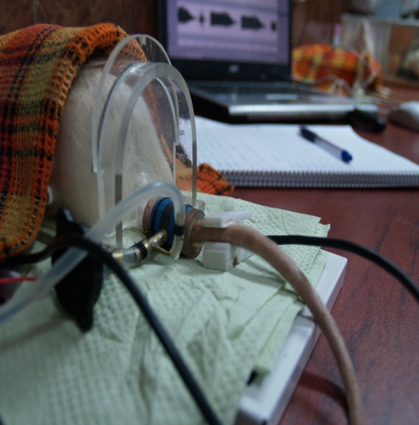
The procedure of a blood pressure measurement in spontaneously hypertensive rats
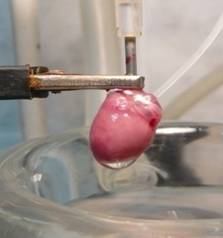
An assessment of cardioprotective, cardiotonic and antiarrhythmic properties of PA in the model of the isolated heart by Langendorf’ method
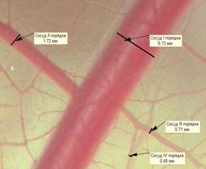
An assessment of the efficiency and the mechanisms of PA action on chicken embryo chorioallantonic membrane vessels
The estimated parameters are the follows:
Total protein,
Albumin,
Total lipid level,
Total cholesterol,
α-cholesterol,
Triglycerides,,
Lipoproteins,
Creatinine,
Creatine kinase,
MV and VV-fractions of creatine kinase,
Lactate dehydrogenase,
ALT and AST,
Lipid peroxidation level, and others.
Histological, histochemical and immunohistochemical studies include:
histological assessment of the presence of ischemic tissues and necrotic areas and morphometric analysis of the affected areas;
an assessment of morphological parameters of elastic and muscle-elastic type blood vessels and vessels of microvasculature in tissues: heart, lungs, pancreas, mesenteric tissue, brain and eye;
histochemical assessment of plasma extravasation in the ischemic tissue;
immunohistological assessment of the following parameters:
the level of an endothelial NO-synthase expression;
the quantity of angiogenesis marker CD31 (PECAM-1);
the quantity of proliferation marker Ki67;
the quantity of proapoptotic marker p53;
the quantity of antiapoptotic protein Mcl-1.
Publications by topic:
Makarov V.G., Makarova M.N., Selezneva A.I., Huttunen O.E., Zabozlaev A.A., Djachuk G.I. Experemental evaluation of antihypertensive activity of new drug valeocor-Q10 in SHR // Phytopharm. 2011. 15th International congress. Book of Nuremberg.Germany 25-27 July 2011. P.72.
Makarov V.G., Makarova M.N., Selezneva A.I., Huttunen O.E., Zabozlaev A.A., Djachuk G.I. Experemental evaluation of antihypertensive activity of new drug valeocor-Q10 in SHR // Phytopharm. 2011. 15th International congress. Book of Nuremberg.Germany 25-27 July 2011. P.72.
Makarova M.N., Selezneva A.I., Makarov V.G. A new model for study of hypo- and hypertensive effects in ovo // In abstract book. The first collaborative meeting on phytomedicine. Monte Verita. Ascona – Switzerland. I 1-13 May 2007. 69.
Makarova M.N., Tesakova S.V., Makarov V.G., Samusenko I.A. Experimental modeling of diabetes mellitus and its complications // Phytopharm. 2008. 12 th International congress. Book of Leiden. Netherlands. 1-4 July 2008. P.71.
Obukhova V.V., Makarova M.N., Khodko S.V., Abrashova V., Sokolova A.P.An investigation of the herbal mixtures influence on the blood coagulation system in a rabbit experimental atherosclerosis model // Phytopharm. 2011. 15th International congress. Book of abstracts. Nuremberg.Germany 25-27 July 2011. P.79-80.
Obukhova V.V., Ivanova S.A., Makarova M.N., Khodko S.V. An investigation of the herbal mixtures influence on the bile composition in a rabbit experimental atherosclerosis model // Phytopharm. 2011. 15th International congress. Book of Nuremberg.Germany 25-27 July 2011. P.80-81
Obukhova V.V., Makarova M.N., Khodko S.V., Abrashova T.V., Sokolova A.P. An investigation of the hypolipidemic activity of herbal mixtures in wistar rats // Phytopharm. 2011. 15th International congress. Book of Nuremberg.Germany 25-27 July 2011. P.78-79.
Obukhova V.V., Makarova M.N., Khodko S.V., Stephanov S.Y. An investigation of the anti- atherosclerotic activity of herbal mixtures in chinchilla rabbits // Phytopharm. 2011. 15th International congress. Book of Nuremberg.Germany 25-27 July 2011. P.82-83
Obukhova V.V., Selezneva A.I., Makarova M.N., Khodko S.V. An investigation of the anti-oxidant activity of herbal mixtures on an experimental hyperlipidemia model in wistar rats // Phytopharm. 2011. 15th International congress. Book of Nuremberg.Germany 25-27 July 2011. P.81-82
Pozharitskaya O.N., Karlina M.V., Shikov A.N., Makarova M.N., Tikchonov V.P. Het- cam assay for vasodilatation effect of nanosystem with taxifolin // In abstract book. The first collaborative meeting on phytomedicine. Monte Verita. Ascona – Switzerland. I 1-13 May 2007. 75
Ryzhenkov V.E., Makarov V.G., Remesova O.V. New plant origin substances: hypolipidemic and hepatoprotective action. // Canad. J. Cardiol., Absts. 4th Intcrnat. Conf on preventive cardiology, Montreal. – 1997. – Vol.13. – Suppl. B. – P. 343 В.
Tesakova S.V., Makarova M.N., Stolaschuk N.V., Makarov V.G. Capillary-protective activity of combination of ascorbic acid and various flavonoids on a model of endothelial dysfunction // Phytopharm. 2007. 11th International congress. Book of abstracts. Leiden. Netherlands. 27-30 June 2007. –P. 74.
Tikchonov V.P., Makarova M.N., Zajtseva M.A., Makarov V.G., Efficacy of (±)-taxifolin from Larix sibirica (Munchh.) Ledeb. on blood pressure in experiments in vivo // GA. 54th Annual Congress on Medicinal Plant research. 2- august – 2 september 2006, Helsinki, Finland. Planta Medica. -2006. –Vol. 72, № 11. –P. 1035.
Ковалева М.А., Селезнева А.И., Макарова М.Н., Макаров В.Г. эффективность убидекаринона при метаболическом синдроме и артериальной гипертензии в эксперименте // Материалы всероссийской молодежной конференции «Фармакологическая коррекция процессов жизнедеятельности. Доклинические и клинические исследования новых лекарственных препаратов» г. Уфа, 8-10 июля 2012 г. – С. 84-87.
Ковалева М.А., Селезнева А.И., Макарова М.Н., Макаров В.Г., Забозлаев А.А., Дьячук Г.И. Эффективность убидекаринона при метаболическом синдроме и артериальной гипертензии в эксперименте // Профилактическая и клиническая медицина. -2012, №4 (45). –С. 81-84.
Макаров В.Г., Макарова М.Н., Проскурина И.А., Богданов А.Н., Сомов Д.В. Методические рекомендации по доклиническому изучению лекарственных средств для коррекции сахарного диабета, ожирения и метаболического синдрома // В кн.: Руководство по проведению доклинических исследований лекарственных средств. Часть первая. –М. Гриф и К, -2012. -944 с.
Макаров В.Г., Рыженков В.Е., Северцева О.В. и др. Гиполипидемическое и антиоксидантное действие концентрата облепихового масла в эксперименте. // Журн. Вопросы биол. мед. и фармацевтич. химии, М. – 1998. – N 3. – С. 42-44.
Макарова М.Н. Биодоступность и метаболизм флавоноидов // Экспериментальная и клиническая фармакология.- 2011. – 74, №6 С.33-40
Макарова М.Н., Макаров В.Г. Молекулярная биология флавоноидов (Химия, биохимия, фармакология). Руководство для врачей // СПб., СПб., из-во «Лема» 2010 г. С. 428.
Макарова М.Н., Макаров В.Г., Линфляндский В.Г. Флавоноиды и заболевания сердечно-сосудистой системы // Вопросы здорового и диетического питания №2-2011.С.53-60.
Макарова М.Н., Селезнева А.И., Макаров В.Г., Дьячук Г.И., Шиков А.Н., Пожарицкая О.Н. Влияние сока аронии черноплодной на артериальное давление экспериментальных животных // Профилактическая и клиническая медицина. -2012, №3 (44). –С. 45-49.
Рыдловская А.В., Гончар И.В., Тесакова С.В., Столащук Н.В., Гущин В.А., Макаров В.Г. Определение влияния противовоспалительного комбинированного растительного препарата Артрофлекс на сигналинг МАР-Киназ JNK1/2, P38 и ERK1/2 и ядерного фактора kB. Мат. межд. конференции «Рецепция и внутриклеточная сигнализация», Пущино, 2007: 333-335.
Рыженков В.Е., Макаров В.Г. Подходы к экспериментальному изучению гиполипидемического и антиатеросклеротического действия новых средств природного происхождения // IX Междунар. съезд “Фитофарм-2005”: матер. съезда. – Санкт-Петербург, 22-25 июня 2005 г. – Санкт-Петербург. – 2005. – С. 623-628.
Рыженков В.Е., Макаров В.Г., Ремезова О.В., Макарова М.Н. Методические рекомендации по изучению гиполипидемического и антиатеросклеротического действия лекарственных средств // В кн.: Руководство по проведению доклинических исследований лекарственных средств. Часть первая. –М. Гриф и К, -2012. -944 с.
Рыженков В.Е., Макаров В.Г., Ремезова О.В., Макарова М.Н. Методические рекомендации по изучению гиполипидемического и антиатеросклеротического действия лекарственных средств // В кн.: Руководство по проведению доклинических исследований лекарственных средств. Часть первая. –М. Гриф и К, -2012. -944 с.
Рыженков В.Е., Ремезова О.В., Макаров В.Г. Методические указания по изучению гиполипидемического и антиатеросклеротического действия фармакологических веществ // В кн. “Руководство по экспериментальному (доклиническому) изучению новых фармакологических веществ / Под ред. Р.У.Хабриева/. Изд. II, перер. и дополн. – Москва: “Медицина”, 2005. – С.452-461.
Рыженков В.Е., Ремезова О.В., Макаров В.Г. Методические указания по изучению гиполипидемического и антиатеросклеротического действия фармакологических веществ // Руководство по экспериментальному (доклиническому) изучению новых фармакологических веществ. М. – 2000. – С. 224-227.
Селезнева А.И., Ковалева М.А., Макарова М.Н., Дьячук Г.И., Макаров В.Г. Моделирование метаболического синдрома у крыс со спонтанной гипертензией // Материалы 2-ой ежегодной научно-практической конференции «Наука о лабораторных животных: современные подходы», Санкт-Петербург, 21-22 декабря 2012 г. – С. 15-16.
Тихонов В.П., Макаров В.Г., Александрова А.Е., Бржетский В.В., Дискаленко О.В. Сравнительная эффективность трех фитокомплексов на развитие индуцированной хориоретинальной дистрофии у кроликов // IX Междунар. съезд “Фитофарм-2005”: матер. съезда. – Санкт-Петербург, 22-25 июня 2005 г. – Санкт-Петербург. – 2005. – С. 352-357.
Патент RU 2520695 C1 Авторы: Пожарицкая О.Н., Шиков А.Н., Макарова М.Н., Макаров В.Г., Фомичев Ю.С. Комплекс биологически активных веществ для лечения и профилактики заболеваний сердечно-сосудистой системы. Опубликовано 27.06.2014. Бюл. № 18.
Авдеева О.И., Макарова М.Н., Афонькина О.В., Макаренко И.Е., Макаров В.Г., Капанадзе Г.Д. Токсикологическая характеристика антигипертензивных средств, влияющих на ренин-ангиотензиновую систему // Биомедицина. -2014, № 2. –С. 99-104.
Селезнева А.И., Макаренко И.Е., Касторнова А.Е., Ходько С.В., Ковалева М.А. Противоишемические свойства метаболических средств в эксперименте // Биомедицина. -2014, № 3. –С. 23-31.
Макаренко И.Е., Ванатиев Г.В., Лукин Ф.Л., Макарова М.Н., Макаров В.Г. Сравнительная характеристика экспериментальных моделей инсульта// Международный вестник ветеринарии. -2014, №4. –С. 104-112.Сравнительная характеристика экспериментальных моделей инсульта
KalatanovaAVGushchin JA, Faustova NM, Vanati GV Makarov,VGModification ofthe modeling methodof reperfusionmyocardial injuryin rats// Proceedings of the IV-th International Congressof VeterinaryPharmacology and Toxicology “Effective andsafe drugsin veterinary medicine.” St.Petersburg, 2016. C. 83-86.
Косман В.М., Демченко Д.В., Пожарицкая О.Н., Шиков А.Н., Селезнева А.И., Макаров В.Г., Макарова М.Н. Кардиопротекторное действие нового функционального пищевого продукта на основе жира лососевых пород рыб с экстрактом травы // Вопросы питания. 2017. № 1. С. 58-63.
Kulyak Yu. O., Gorodetskaya E.A., Kalenikova E.I., Makarova M.N., Pozharitskaya O.N., Medvedev O.S. Evaluation of cardioprotective efficacy of innovative dosage form of ubiqinol for intravenous administration // Éksperimentalnaya i Klinicheskaya Farmakologiya. – 2018. – Vol.81(4). – P. 8-11. ABCTRACT. Cardioprotective efficacy of innovative dosage form of ubiqinol for intravenous administration was investigated on the experimental myocardial infarction model in rats. According to the assessment made on the 21st day, intravenous administration of the drug injected in 10 min after coronary artery occlusion (10 mg/kg) significantly reduced the size of aneurysm (18.36%, p < 0.05) and prevented from the development of myocardial hypertrophy of the left ventricle. The group of animals receiving the drug showed the inverse correlation (r = –0.6716) between the concentration of CoQ10 in the myocardium and the thickness of the interventricular septum, which underlines its role in postinfarction damage limitation [Full text is available in Russian].